Laboratory Equipment & Techniques for Spider Identification

Laboratory Equipment & Techniques
Preserving Spiders
Live spiders may be immobilised by freezing them in a refrigerator before transferring them into specimen tubes. Spider specimens should be preserved in 70-80% ethanol (ethyl alcohol) and stored in glass tubes or jars with tightly fitted caps so that the risk of evaporation over time may be minimised. To prevent the leakage or evaporation of ethanol, the caps may be sealed with paraffin wax or Parafilm. A more reliable but expensive option is to use “Polyseal” screw cap vials. These come with an interior polyethylene liner or cone projecting into the mouth of the vial. For double protection and easy checks and maintenance, many museums make it a standard practice to immerse specimens in ethanol within glass tubes plugged with cotton wool which are then placed in larger jars filled with ethanol.
Each specimen should be accompanied by a label that includes the date and locality of collection (with GPS reading if available), and the name of the collector. Where possible, the name and sex of the spider should also be included.
For temporary storage, labels may be written in pencil. For longer-term or permanent storage, labels should be written with Indian ink in technical drawing pens, or with alcohol-proof archival ink in disposable “Pigma Micron” pens. Alternatively, labels may be mass-produced speedily using a laser printer and heating the printed sheet in an oven (around 100°C for 10-15 minutes) to melt and embed the carbon particles of the printed words onto the paper. Ordinary ink or bubble jet should not be used as the ink of these printers will dissolve in ethanol. The paper should ideally be acid-free or of archival quality, weighing 250 GSM. Ordinary lightweight printing paper should be avoided as labels made of such paper can disintegrate in ethanol over time.
Laboratory Equipment & Techniques
Examining Spiders
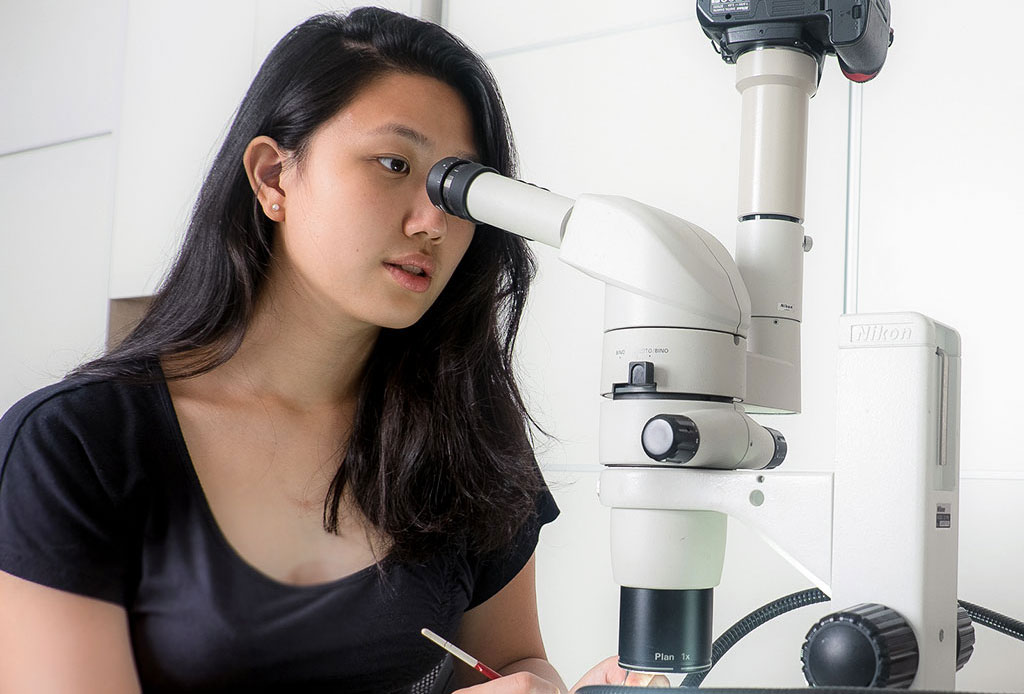
Identification of spiders requires examination of the spiders’ genitalia and other minute morphological structures. For this purpose, a stereoscopic dissecting microscope with a magnification of 20X to 80X will be useful. If a laboratory fibre optic LED light source is not available, sufficient illumination can be obtained from a bright halogen table lamp with a gooseneck.
A specimen should be examined while it is submerged under 75% ethanol in a watch glass or paint palette. The ethanol may be slowly dispensed with a fine-tipped squeeze bottle.


Specimens may be handled with a pair of fine forceps used by watch repairers, and, if necessary, used in combination with fine watercolour paint brushes. Fine watercolour paint brushes can also be useful in brushing away hairs, food remains and other debris from the body to provide a less obscured view of some of the finer morphological details.
Dissection of small body parts can be done with a fine entomological pin mounted on a used syringe or any other form of cylindrical holder. The specimen may be manipulated and held in place at the desired orientation for microscopic examination or photomicroscopy in a bedding of fine sand, fine laboratory glass beads, a layer of hand sanitiser gel, or personal lubricant jelly.
Laboratory Equipment & Techniques
Identifying Spiders
The first step in spider identification is to learn to place them in their respective families. For this purpose, the identification key in Christa Deeleman-Reinhold’s 2001 book will be most useful for South East Asian spiders. Read more in How to Identify Spiders.

This article is adapted from the Laboratory Techniques and Equipment chapter in Spiders of Brunei Darussalam by Joseph K. H. Koh and Leong Tzi Ming, 2013.


